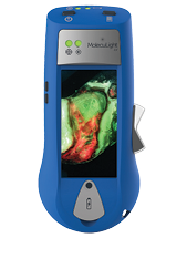
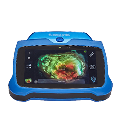
Clinical Image Gallery of Fluorescence Images
Below are a series of images/videos highlighting how the MolecuLight i:X helped clinicians detect fluorescent bacteria and measure wounds to facilitate evidence-based clinician decision making.
Media List
Diabetic Foot Ulcer, Heel
IMAGE
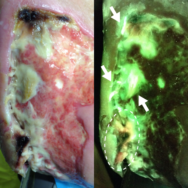
FL-image revealed both cyan fluorescing bacteria which is indicative of Pseudomonas aeruginosa (arrows) and red fluorescing bacteria

Figure 1: ST-image
Figure 2: FL-image
Diabetic Foot Ulcer, Heel
FL-image revealed both cyan fluorescing bacteria which is indicative of Pseudomonas aeruginosa (arrows) and red fluorescing bacteria (circled) which guided swabbing for accurate microbiology results.

Anatomical Location:
Left Foot, Heel
Microbiology Results:
Curettage samples confirmed heavy growth of Pseudomonas aeruginosa and light growth of Staphylococcus aureus in this wound
Image/Video Provided By:
Ron Linden, MD, Judy Dan Research & Treatment Centre, Toronto, ON, Canada
Case ID:
MolecuLight Clinical Case 0044
Burn Injury, Leg
IMAGE
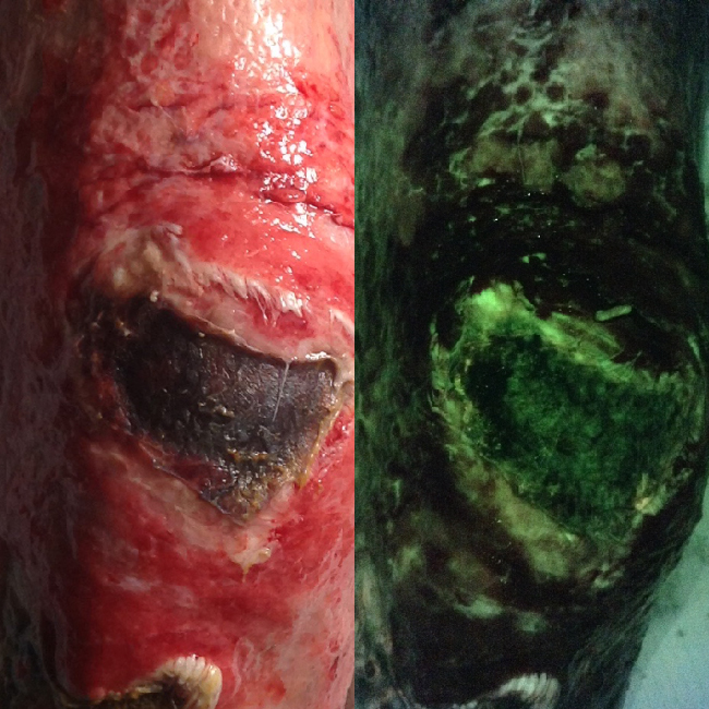
Absence of red fluorescence in FL-image suggests no significant bioburden which provided clinician with confidence

Figure 1: ST-image
Figure 2: FL-image
Burn Injury, Leg
Absence of red fluorescence in FL-image suggests no significant bioburden which provided clinician with confidence to proceed with a skin graft procedure.
Anatomical Location:
Right Leg
Microbiology Results:
Earlier microbiology reports confirmed negative for bacteria (sample taken days prior)
Image/Video Provided By:
Lt Col. Steven Jeffery, The Royal Centre for Defence Medicine, Birmingham, UK
Case ID:
MolecuLight Clinical Case 0055
Pectoral Muscle
IMAGE
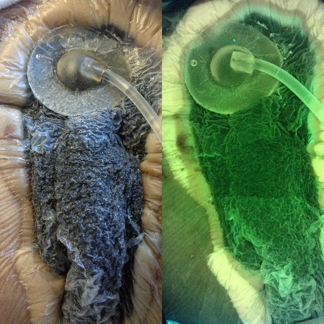
Absence of red fluorescence in FL-image suggests no bacteria (later confirmed via swabs); clinician decided

Figure 1: ST-image
Figure 2: FL-image
Pectoral Muscle
Absence of red fluorescence in FL-image suggests no bacteria (later confirmed via swabs); clinician decided to leave wound bed undisturbed for an additional 24 hours.
Anatomical Location:
Pectoral
Microbiology Results:
Swabs results, taken during next dressing change, were negative for bacteria
Image/Video Provided By:
Rose Raizman, RN-EC, MSc, Scarborough & Rouge Hospital, Toronto, ON, Canada
Case ID:
MolecuLight Clinical Case 0041
Venous Leg Ulcer
IMAGE
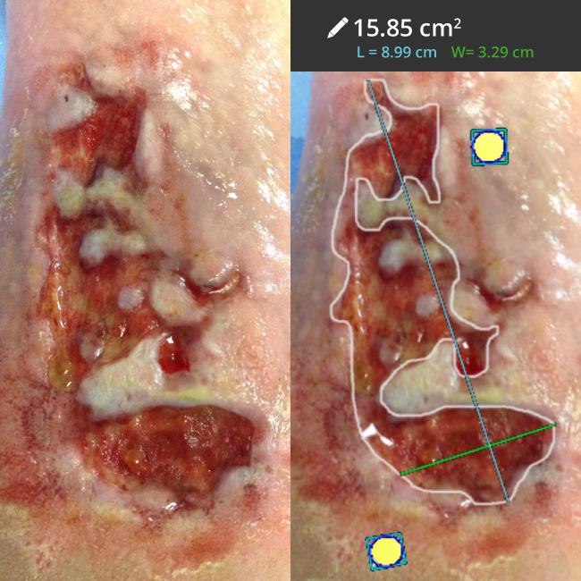
Wound measurement with the MolecuLight i:X allowed for more accurate measurement of this wound with

Figure 1: ST-image
Figure 2: ST-image with Wound Measurement
Venous Leg Ulcer
Wound measurement with the MolecuLight i:X allowed for more accurate measurement of this wound with an irregular wound border.

Anatomical Location:
Left Leg, Calf
Image/Video Provided By:
Rose Raizman, RN-EC, MSc, Scarborough & Rouge Hospital, Toronto, ON, Canada
Case ID:
MolecuLight Clinical Case 0018
Surgical Site Infection, Amputation
VIDEO
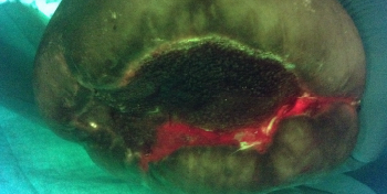
The clinician used the MolecuLight i:X in Fluorescence Imaging Mode to visualize bacterial contamination of
Surgical Site Infection, Amputation
The clinician used the MolecuLight i:X in Fluorescence Imaging Mode to visualize bacterial contamination of a limb amputation in real-time. Red fluorescence indicates presence of bacteria at the surgical site. As a result, further wash-out was performed prior to closure.
Anatomical Location:
Leg
Microbiology Results:
Swabs confirmed E. coli and P. mirabilis
Image/Video Provided By:
Lt Col. Steven Jeffery, The Royal Centre for Defence Medicine, Birmingham, UK
Case ID:
MolecuLight Clinical Case 0014
Surgical Site Infection, Abdomen
VIDEO
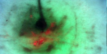
The clinician used the MolecuLight i:X in Fluorescence Imaging Mode to target cleaning to areas
Surgical Site Infection, Abdomen
The clinician used the MolecuLight i:X in Fluorescence Imaging Mode to target cleaning to areas of red fluorescence (red indicates presence of bacteria) throughout wound healing of a patient’s abdominal surgery.

Anatomical Location:
Abdomen
Image/Video Provided By:
Rose Raizman, RN-EC, MSc, Scarborough & Rouge Hospital, Toronto, ON, Canada
Case ID:
MolecuLight Clinical Case 0010
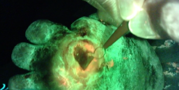
Diabetic Foot Ulcer
VIDEO
The clinician used the MolecuLight i:X to guide debridement of a heavy callus on a
Diabetic Foot Ulcer
The clinician used the MolecuLight i:X to guide debridement of a heavy callus on a diabetic foot ulcer. As the wound is debrided, the MolecuLight i:X reveals a red fluorescent signal, indicating the presence of bacteria in the wound.
Anatomical Location:
Left Foot
Image/Video Provided By:
Rose Raizman, RN-EC, MSc, Scarborough & Rouge Hospital, Toronto, ON, Canada
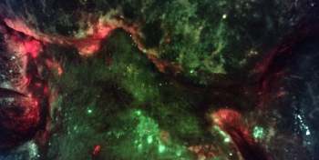
Venous Leg Ulcer, Obesity-associated Lymphedema – Part 1
VIDEO
By incorporating MolecuLight i:X into the cleaning protocol, the clinician was able to visualize moderate/heavy
Venous Leg Ulcer, Obesity-associated Lymphedema – Part 1
By incorporating MolecuLight i:X into the cleaning protocol, the clinician was able to visualize moderate/heavy bacterial load (red fluorescence) in the creases and at the periphery of the wound in real-time to guide cleaning.
Anatomical Location:
Leg
Image/Video Provided By:
Rose Raizman, RN-EC, MSc, Scarborough & Rouge Hospital, Toronto, ON, Canada
Case ID:
MolecuLight Clinical Case 0017
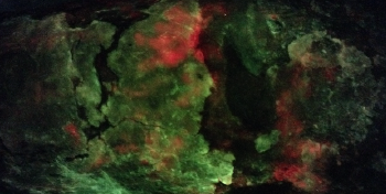
Venous Leg Ulcer, Secondary Cellulitis
VIDEO
See the progress of a venous leg ulcer with secondary cellulitis over the course of
Venous Leg Ulcer, Secondary Cellulitis
See the progress of a venous leg ulcer with secondary cellulitis over the course of a few weeks. The clinician used the MolecuLight i:X to visualize bacteria (which fluoresce red) to target wound cleaning.
Anatomical Location:
Leg
Image/Video Provided By:
Rose Raizman, RN-EC, MSc, Scarborough & Rouge Hospital, Toronto, ON, Canada
Case ID:
MolecuLight Clinical Case 0037
