Case Studies
Below are a series of case studies highlighting how the MolecuLight i:X easily integrates into wound care protocols and guides clinicians in their wound care practice.
Featured Case Study
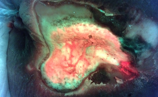
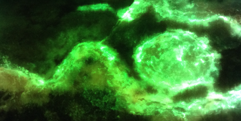
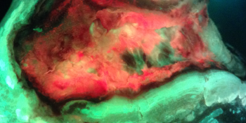
Real-Time Detection of Asymptomatic Bioburden in Wounds
Use of the MolecuLight i:X to image for bacterial fluorescence, in conjunction with clinical signs & symptoms, adds an additional bacterial-specific piece of information that can be captured and considered in real-time.
See Full Case StudyCase Studies by Group
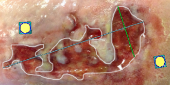
Visualize fluorescent bacteria and measure wound surface area in real-time to understand the...
See Assessment cases >
Visualize fluorescent bacteria and measure wound surface area in real-time to understand the status of the wound more fully.
Wound Healing Cost Savings with MolecuLight i:X Case Study
Read More >
Allows clinicians to focus cleaning in areas where fluorescent bacteria are located and optimize...
See Cleaning cases >
Allows clinicians to focus cleaning in areas where fluorescent bacteria are located and optimize wound bed preparation.
Guides more efficient and targeted debridement.
See Debridement cases >
Guides more efficient and targeted debridement.
Guides where to sample; 50% more accurate swabbing compared to the Levine Technique.
See Sampling cases >
Guides where to sample; 50% more accurate swabbing compared to the Levine Technique.
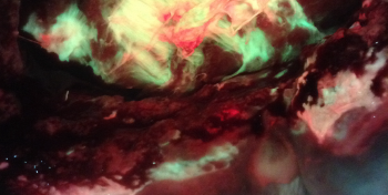
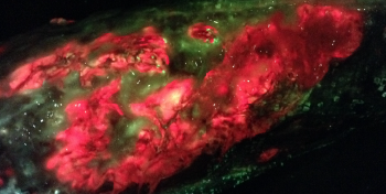
Visualizing Pseudomonas Aeruginosa and Optimized Sampling with the MolecuLight i:X
Read More >
Provides objective visual documentation of the presence of fluorescent bacteria and the surface...
See Documentation cases >
Provides objective visual documentation of the presence of fluorescent bacteria and the surface area of the wound.
Comparing fluorescent bacteria and wound surface area at each visit may provide real-time objective...
See Treatment Selection cases >
Comparing fluorescent bacteria and wound surface area at each visit may provide real-time objective feedback on impact of treatment plan.
Supports more responsible antibiotic decision making and selection.
See Antibiotic Stewardship cases >
Supports more responsible antibiotic decision making and selection.
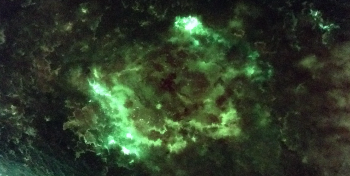
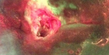
Point of Care Identification of Pseudomonas Aeruginosa Case Study
Read More >
Patients easily see and understand why a clinician is taking certain action to clean, debride...
See Patient Engagement cases >
Patients easily see and understand why a clinician is taking certain action to clean, debride and treat a wound in a specific way.
References
- Wu YC, Smith M, Chu A, Lindvere-Teene L, Starr D, Tapang K, Wong O, Linden R, DaCosta RS. Handheld fluorescence imaging device detects subclinical wound infection in an asymptomatic patient with chronic diabetic foot ulcer: a case report. International Wound Journal, 2016, 13(4), 449-53. doi: 10.1111/iwj.12451.
- DaCosta RS, Kulbatski I, Lindvere-Teene L, Starr D, Blackmore K, Silver JI, Opoku J, Wu YC, Medeiros PJ, Xu W, Xu L, Wilson BC, Rosen C, Linden R. Point-of-care autofluorescence imaging for real-time sampling and treatment guidance of bioburden in chronic wounds: first-in-human results. PLOS ONE, 2015, 10(2). doi: 10.1371/journal.pone.0116623.
- Ottolino-Perry K, Chamma E, Blackmore KM, Lindvere-Teene L, Starr D, Tapang K, Rosen CF, Pitcher B, Panzarella T, Linden R, DaCosta RS. Improved detection of clinically relevant wound bacteria using autofluorescence image-guided sampling in diabetic foot ulcers. International Wound Journal, 2017, 14(5), 833-841. doi: 10.1111/iwj.12717.
- Rennie MY, Lindvere-Teene L, Tapang K, Linden R. Point-of-care fluorescence imaging predicts the presence of pathogenic bacteria in wounds: a clinical study. Journal of Wound Care, 2017, 26(8), 452-460. doi: 10.12968/jowc.2017.26.8.452.
- Raizman R. Point-of-care fluorescence imaging device guides care and patient education in obese patients with surgical site infections. Presented at CAWC 2016. Proceedings of the Annual Canadian Association of Wound Care Conference; 2016 Nov 3-6, Niagara Falls, ON, Canada.
- Jeffery S. Utility of point-of-care autofluorescence imaging device in successful closure of major limb amputations – a case study. Presented at MHSRS 2016. Proceedings of the Military Health System Research Symposium; 2016 Aug 15-18; Kissimmee, FL, USA.
- Dunham D, Teene L. Objective wound measurement software on a point-of-care, hand-held fluorescence imaging device: verification of measurement accuracy and repeatability. Presented at EWMA 2018. Proceedings of the Annual European Wound Management Association Conference; 2018 May 9-11; Krakow, Poland.
- Wu YC, Kulbatski I, Medeiros PJ, Maeda A, Bu J, Xu L, Chen Y, DaCosta RS. Autofluorescence imaging device for real-time detection and tracking of pathogenic bacteria in a mouse skin wound model: preclinical feasibility studies. Journal of Biomedical Optics, 2014, 19(8). doi: 10.1117/1.JBO.19.8.085002.
- Raizman R, DaCosta RS. Handheld real-time fluorescence imaging of bacteria guides treatment selection and timing of dressing changes in inpatients undergoing negative pressure wound therapy. Presented at IWH 2016. Proceedings of the Innovations in Wound Healing Conference; 2016 Dec 8-11, Key Largo, FL, USA.
- Landis S, Rennie MY, Blumenthal E, Jeffery S. Use of fluorescence imaging in visualizing bacteria in chronic ulcers and traumatic soft tissue damage. Presented at AMSUS 2016. Proceedings of the Annual Meeting of the Society of Federal Health Professionals; 2016 Nov 29-Dec 2; National Harbor, MD, USA.


