Reliability in microbiology wound sampling
In the article ‘Improved detection of clinically relevant wound bacteria using autofluorescence image-guided sampling in diabetic foot ulcers’ recently published (March 2017) in International Wound Journal, Ottolino-Perry et al. present data from a clinical trial highlighting how the conventional Levine method of sampling may need to be reconsidered as an accepted standard for its reliability in microbiology wound sampling.
It is accepted that for patients with a suspected wound infection, wound cultures are an important part in clinically diagnosing the infection, identifying the specific organisms, and determining the number of organisms present. This information helps to guide appropriate antibiotic selection and is crucial in minimizing antibiotic-resistant infections. Therefore, obtaining an accurate sample representative of the microbiological profile of a wound is imperative during the diagnosis process for the optimum management of wound infections.
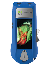
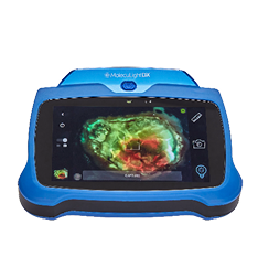
Ottolino-Perry et al. demonstrate that point-of-care fluorescence image-guided swabbing more accurately identified the presence of moderate and/or heavy bacterial loads compared with the Levine technique in diabetic foot ulcers. Importantly, fluorescence imaging detected and identified pathogens (S. aureus and E. coli) that were otherwise missed during routine clinical signs and symptoms assessment alone. Therefore, an accurate and representative sampling of the wound is critical for appropriate antimicrobial treatment decisions, as the sensitivity of different species to antibiotic regimens can vary greatly. The authors show that fluorescence image-guided swabbing was easy to perform and maximized the effectiveness of wound sampling, with no significant impact on clinical workflow. The study confirmed that fluorescence imaging can help clinicians overcome the limitations of traditional sampling methods by better identifying specific wound areas containing clinically significant bacteria in real time. This helped maximize the effectiveness of sampling of treatment-relevant pathogens in diabetic foot ulcers.
Click here to read full article
