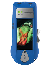
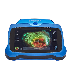
The clinical signs and symptoms of infection in acute and chronic wounds are unreliable. Similarly, swab cultures are inaccurate in this population. Tissue biopsies and polymerase chain reaction (PCR) are more accurate but the results require several days to obtain. As a result, the clinician is forced to treat patients empirically. This has led to the overuse of antibiotics and the failure of advanced therapies due to unrecognized infection. To address this problem a point-of-care diagnostic was developed to identify bacteria in acute and chronic wounds. The MolecuLight procedure (MolecuLight i:X) exposes the wound bed to violet light at 405 nm. Bacterial fluorophores absorb the light. In turn they fluoresce at specific wavelengths: porphyrins (red) and pyoverdines (cyan). The device detects bacteria in the wound bed at a level greater than 104 by measuring the amounts of red and or cyan fluorescence. A robust body of literature has demonstrated that elevated bacterial levels impede wound healing. The MolecuLight i:X can detect elevated bacteria burden in a wound allowing the clinician to address the infection. In addition, the device can guide advanced wound therapies such as antibiofilm agents, negative wound pressure therapy and preparation of the wound bed for grafting.
