ABSTRACT
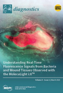
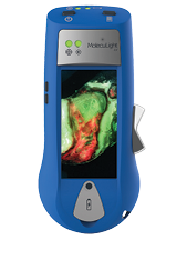
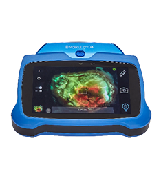
The persistent presence of pathogenic bacteria is one of the main obstacles to wound healing. Detection of wound bacteria relies on sampling methods, which delay confirmation by several days. However, a novel handheld fluorescence imaging device has recently enabled real-time detection of bacteria in wounds based on their intrinsic fluorescence characteristics, which differ from those of background tissues. This device illuminates the wound with violet (405 nm) light, causing tissues and bacteria to produce endogenous, characteristic fluorescence signals that are filtered and displayed on the device screen in real-time. The resulting images allow for rapid assessment and documentation of the presence, location, and extent of fluorescent bacteria at moderate-to-heavy loads. This information has been shown to assist in wound assessment and guide patient-specific treatment plans. However, proper image interpretation is essential to assessing this information. To properly identify regions of bacterial fluorescence, users must understand: (1) Fluorescence signals from tissues (e.g., wound tissues, tendon, bone) and fluids (e.g., blood, pus); (2) fluorescence signals from bacteria (red or cyan); (3) the rationale for varying hues of both tissue and bacterial fluorescence; (4) image artifacts that can occur; and (5) some potentially confounding signals from non-biological materials (e.g., fluorescent cleansing solutions). Therefore, this tutorial provides clinicians with a rationale for identifying common wound fluorescence characteristics. Clinical examples are intended to help clinicians with image interpretation—with a focus on image artifacts and potential confounders of image interpretation—and suggestions of how to overcome such challenges when imaging wounds in clinical practice.
