Abstract
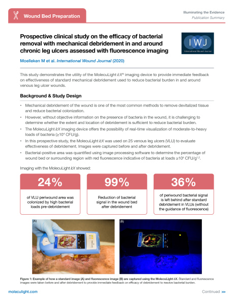
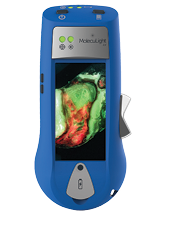
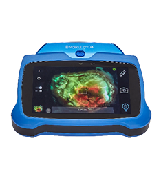
Bacterial colonisation in wounds delays healing, mandating regular bacterial removal through cleaning and debridement. Real-time monitoring of the efficacy of mechanical debridement has recently become possible through fluorescence imaging (MolecuLight i:X®). Red fluorescence, endogenously produced during bacterial metabolism, indicates regions contaminated with live bacteria (>104 CFU/g). In this prospective study, conventional and fluorescence photos were taken of 25 venous leg ulcers before and after mechanical debridement, without use of antiseptics. Images were digitally segmented into wound bed and the periwound regions (up to 1.5 cm outside bed) and pixel intensity of red fluorescence evaluated to compute bacterial area. Pre-debridement, bacterial fluorescence comprised 10.4% of wound beds and larger percentages of the periwound area (~25%). Average bacterial reduction observed in the wound bed after a single mechanical debridement was 99.4% (p<0.001), yet periwound bacterial reduction was only 64.3%. On average, across bed and periwound, a single mechanical debridement left behind 29% of bacterial fluorescence positive tissue regions. Our results show the substantial effect that safe, inexpensive, mechanical debridement can have
on bacterial load of venous ulcers without antiseptic use. Fluorescence imaging can localise bacterial colonised areas and showed persistent periwound bacteria post-debridement. Fluorescence-targeted debridement can be used quickly and easily in daily practice.
KEYWORDS
bacteria, chronic leg ulcers, fluorescence, MolecuLight, wound debridement
