ABSTRACT
BACKGROUND: Partial and full thickness burns require surgical treatment, such as early débridement and skin transplantation in MEEK/MESH technique or further reconstructive surgery. Infections of burns or transplanted areas limit surgical success and increase patient mortality. For split-thickness grafts in MEEK technique a superficial silk is applied as a protective on-top dressing, whereas in MESH technique fatty gauze and foam are used as standard protective covers over five to seven days. However, wound occlusion by both materials provides the soil for growth of microorganisms. The timely identification of impending infections is necessary to initiate early removal in order to safe and preserve skin grafts. Early identification of infections and removal of foreign material should therefore be attempted.
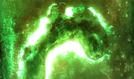
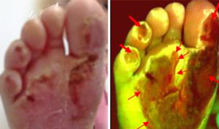
MATERIAL AND METHODS: Burn wounds treated with split-thickness skin grafts processed by MEEK/MESH technique and covered with silk or foam overlayers were analyzed for signs of bacterial infection using the MolecuLight i:X™ device. In addition, swaps for microbiological analysis where taken from fluorescent areas and correlated with florescent image results.
RESULTS: We examined burn wounds (n = 14) of three different intensive care patients. The MolecuLight i:X™ camera showed a strong colonization of the transplanted areas and foreign materials, that were in line with microbiological analysis findings. The representation of the excitation load showed high values in the foreign materials. The take rate of MEEK-transplants was 90 % compared to MESH-transplanted with about 60 %. The positive predictive value was 81.8 % for detection of a wound infection with autofluorescence. The negative predictive value was 90.3 % with a sensitivity of 86.7 % and a specificity of 87.5 %.
CONCLUSION: The representation of the fluorescence exciter load shows high concentrations of pathogens both in the MEEK silk layer as well as in foam linkers. Overall split-thickness grafts according to the MEEK technique showed a higher healing rate compared to MESH technique. Screening of burns wounds with autofluorescence imaging can be helpful for an additive wound assessment. Split-thickness graft covers should be applied only for a minimum time period required to ensure stable grafting.