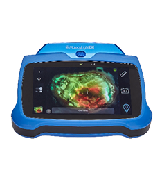
To estimate comparative healing rates and decision-making associated with the use of bacterial autofluorescence imaging in the management of diabetic foot ulcers (DFUs).
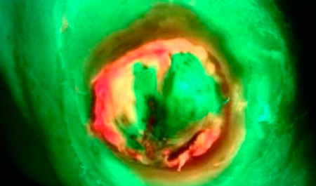
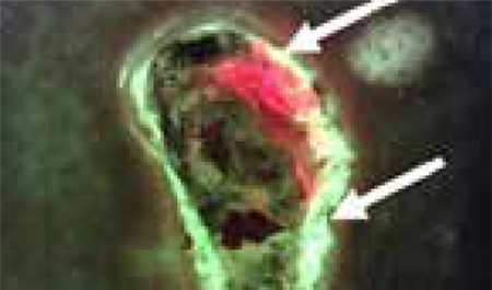
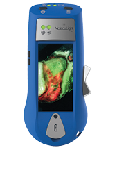
This is a single-center (multidisciplinary outpatient clinic), prospective pilot, randomized controlled trial (RCT) in patients with an active DFU and no suspected clinical infection. Consenting patients were randomly assigned 1:1 to either treatment as usual informed by autofluorescence imaging (intervention), or treatment as usual alone (control). The primary outcome was the proportion of ulcers healed at 12 weeks by blinded assessment. Secondary outcomes included wound area reduction at 4 and 12 weeks, patient quality of life, and change in management decisions after autofluorescence imaging.
Between November 2017 and November 2019, 56 patients were randomly assigned to the control or intervention group. The proportion of ulcers healed at 12 weeks in the autofluorescence arm was 45% (n = 13 of 29) vs. 22% (n = 6 of 27) in the control arm. Wound area reduction was 40.4% (autofluorescence) vs. 38.6% (control) at 4 weeks and 91.3% (autofluorescence) vs. 72.8% (control) at 12 weeks. Wound debridement was the most common intervention in wounds with positive autofluorescence imaging. There was a stepwise trend in healing favoring those with negative autofluorescence imaging, followed by those with positive autofluorescence who had intervention, and finally those with positive autofluorescence with no intervention.
In the first RCT, to our knowledge, assessing the use of autofluorescence imaging in DFU management, our results suggest that a powered RCT is feasible and justified. Autofluorescence may be valuable in addition to standard care in the management of DFU.
