
Fluorescence imaging using MolecuLight provides a more accurate and relevant microbiological profile that guides optimal wound sampling compared to clinical judgment
Fluorescence imaging using MolecuLight provides a more accurate and relevant microbiological profile that guides optimal wound sampling compared to clinical judgment
In a clinical trial with 412 punch biopsies, only one resulted in an adverse event (abscess)
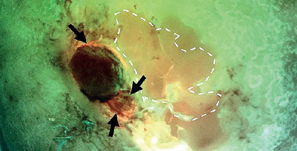
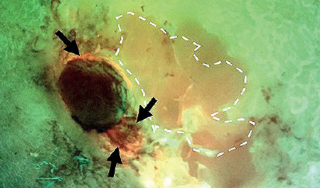
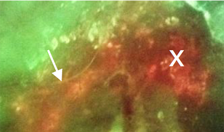
Fluorescence images revealed presence of bacteria at site of biopsy prior to abscess development
Bacterial load was confirmed as 106 CFU/g from the biopsy
The PPV of red fluorescence on MolecuLight i:X images was 100%, regardless of sampling method, analysis technique, or study site
Fluorescence guidance, in combination with subsurface sampling techniques, could eliminate the risk of false negative wound sampling
Fluorescence imaging information could influence treatment decisions at the point of care
Fluorescence imaging of diabetic foot ulcers had superior accuracy (78%) in detecting the presence of clinically significant bacteria than standard practice (Levine swabbing, 52%; p=0.048)
Higher bacterial loads were detected from wound regions positive for bacterial fluorescence compared to regions sampled based on CSS alone


MolecuLight is SOC ll®
Type l Accredited:

MolecuLight awarded
Premier’s Technology
Breakthrough Designation

MolecuLight i:X
is designated as a:

MolecuLight included
in ISWCAP Consensus
Guidelines:
MolecuLight is a proud
corporate member of:
©2025
The MolecuLight® i:X and MolecuLightDX™ Imaging Devices are approved by Health Canada for sale in Canada and has CE marking for sale in the European Union.
The MolecuLight™ i:X and DX Imaging Devices have received FDA clearance.