See what you’re missing with MolecuLight i:X
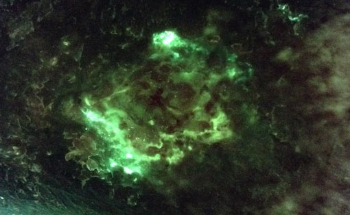
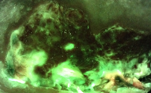
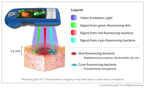
The MolecuLight i:X is a handheld fluorescence imaging device that provides instant visual detection and documentation of potentially harmful bacteria in wounds that would otherwise be invisible.1 The MolecuLight i:X emits a precise wavelength of safe violet light, which interacts with the wound tissue and bacteria causing the wound and surrounding skin to emit a green fluorescence (i.e. collagen) and potentially harmful bacteria to emit a red fluorescence (i.e. porphyrins).1 In real-time, MolecuLight i:X captures these red and green fluorescence signals using specialized optical components to filter out the violet light, and displays the resultant image immediately on the display screen (FL-image).1,2 The MolecuLight i:X is precisely calibrated to detect fluorescent bacteria at levels of ≥ 104 CFU/g on a quantitative scale or predominantly moderate to heavy growth on a semi-quantitative scale.3
Which types of bacteria can be detected?
Pre-clinical and clinical studies have shown that the MolecuLight i:X can detect red fluorescence from Gram positive, Gram negative, aerobic and anaerobic bacterial species1,5.
Pre-clinical research has demonstrated the following species can produce red fluorescence detectable by the MolecuLight i:X in vitro5 (see list below). However, there are many bacterial species not tested here that may also produce red fluorescence6.
- Staphylococcus aureus
- Staphylococcus epidermidis
- Staphylococcus capitis
- Staphylococcus lugdunensis
- Pseudomonas aeruginosa
- Pseudomonas putida
- Escherichia coli
- Corynebacterium striatum
- Proteus mirabilis
- Proteus vulgaris
- Enterobacter cloacae
- Serratia marcescens
- Acinetobacter baumannii
- Klebsiella pneumoniae
- Klebsiella oxytoca
- Morganella morganii
- Propionibacterium acnes
- Stenotrophonomas maltophilia
- Bacteroides fragilis
- Aeromonas hydrophilia
- Alcaligenes faecalis
- Bacillus cereus
- Citrobacter koseri
- Citrobacter freundii
- Clostridium perfringens
- Listeria monocytogenes
- Peptostreptococcus anaerobius
- Veillonella parvula