ABSTRACT
Background:
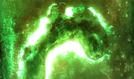
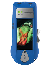
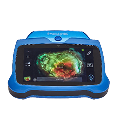
Mechanisms for military injury have evolved in the past century. Debridement is the gold standard for preparing a clean wound bed, decreasing the bacterial load, and reducing the likelihood of infection. However, bacteria may continue to linger in these wounds. The MolecuLight i:X Imaging Device uses the principle of autofluorescence to detect bacteria under violet light. Thus, visualizing bacteria will not only guide clinicians in their management of the wound but it will also serve as a means of evaluating debridement efforts. We hope to improve traumatic wound management by targeting debridement and assessing its quality.
Methods:
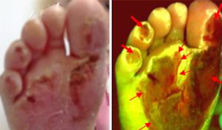
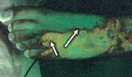
We describe the use of the MolecuLight i:X to photograph wounds under standard and violet light in three patients. Images were captured before and after debridement. Microbiology swabs were collected to correlate the bacteria found in the images to the wounds.
Results:
The post-debridement images demonstrate a marked decrease or complete removal of bacteria. The microbiology swabs confirmed the pre-debridement presence of bacteria.
Conclusions:
The MolecuLight i:X shows promise for debridement evaluation. The use of the MolecuLight i:X may reduce the likelihood of infection, thus having positive implications for military and trauma wounds.
