ABSTRACT
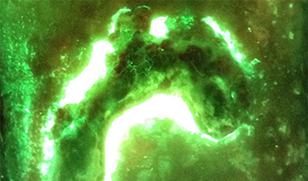
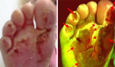
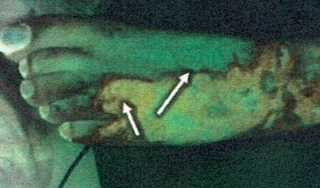
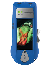
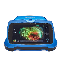
The MolecuLight i:X Imaging Device is a portable, noninvasive, real-time camera used to visualize the bacterial load in a wound. It uses violet light illumination and a dual bandpass optical filter to capture the fluorescence of endogenous structures in the tissue matrix and harmful bacteria. The MolecuLight i:X captures images of wounds and highlights potentially detrimental levels of bacteria. This is an initial evaluation of using the MolecuLight i:X camera in the management of burns to demonstrate the following: the ability of the device to guide clinicians in their management of the burn (i.e. detect, identify, and specify swabbing locations). Burn wounds were photographed under standard light and violet light illumination to compare presentations of obvious infection signs and symptoms. Microbiology swab samples were obtained to correlate any bacterial presence to the images. The fluorescence images were used to guide swabs to where the bacteria were congregating. Twenty patients were imaged. Four patients did not have bacterial contamination based on their images and swab results. Sixteen patients showed growth of Staphylococcus aureus, Pseudomonas aeruginosa, or other bacteria. Nine of the patients, by definition, had infections. These findings were correlated with the typical signs and symptoms of infection, the fluorescence images, and the microbiology results. The efficacy of the MolecuLight i:X is evident due to the microbiology results correlating to the images. Further research is being done to test the device in terms of being an early intervention tool. With these early results and guidance of swab samples, the MolecuLight i:X may be able to detect bacterial load before an infection and subsequent graft failure, thereby shortening lengths of hospital stay and improving overall healing.
