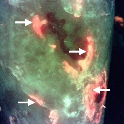
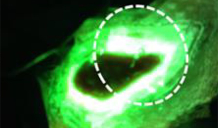
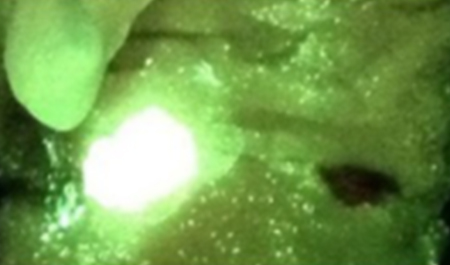
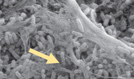
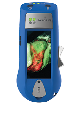
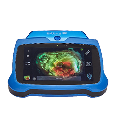
Pseudomonas aeruginosa (PA) is a common bacterial pathogen in chronic wounds known for its propensity to form biofilms and evade conventional treatment methods. Early detection of PA in wounds is critical to the mitigation of more severe wound outcomes. Point-of-care bacterial fluorescence imaging (MolecuLight i:X) illuminates wounds with safe, violet light, triggering the production of cyan fluorescence from PA. A prospective single blind clinical study was conducted to determine the positive predictive value (PPV) of cyan fluorescence for the detection of PA in wounds. Bacterial fluorescence using the MolecuLight i:X imaging device revealed cyan fluorescence signal in 28 chronic wounds, including venous leg ulcers, surgical wounds, diabetic foot ulcers and other wound types. To correlate the cyan signal to the presence of PA, wound regions positive for cyan fluorescence were sampled via curettage. A semi-quantitative culture analysis of curettage samples confirmed the presence of PA in 26/28 wounds, resulting in a PPV of 92.9%. The bacterial load of PA from cyan-positive regions ranged from light to heavy. Less than 20% of wounds that were positive for PA exhibited the classic symptoms of PA infection. These findings suggest that cyan detected on fluorescence images can be used to reliably predict bacteria, specifically PA at the point-of-care.
Back to All Clinical Evidence
Species Detected/Biofilm, Chronic Wounds
Rapid Diagnosis of Pseudomonas aeruginosa in Wounds with Point-of-Care Fluorescence Imaging
Pseudomonas aeruginosa, a common bacterial pathogen in chronic wounds, is challenging to detect by standard assessment of clinical signs and symptoms
Cyan detected on MolecuLight i:X fluorescence images, can be used to reliably predict Pseudomonas aeruginosa at the point-of-care, with a PPV of 93% (confirmed by microbiological analysis)
