Back to All Clinical Evidence
Evidence Category:
Species Detected/Biofilm

Species Detected/Biofilm

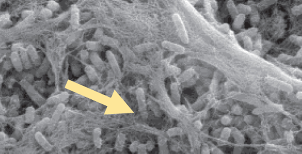
Efficient, accurate diagnosis of cellulitis critical to avoid costly complications

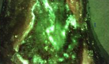
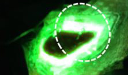
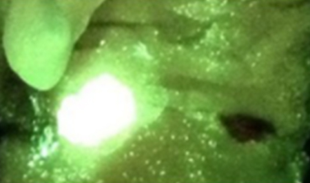
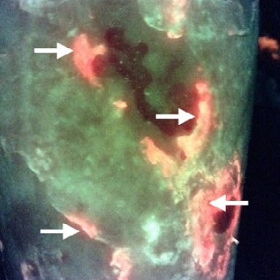
Unique pattern of red fluorescence extending beyond the wound was observed in all cases of wound-related cellulitis

Fluorescence imaging aided in the early diagnosis of wound-related cellulitis, resulting in the initiation of oral antibiotics without delay, avoiding progressive cellulitis


Pseudomonas aeruginosa, a common bacterial pathogen in chronic wounds, is challenging to detect by standard assessment of clinical signs and symptoms

Cyan detected on MolecuLight i:X fluorescence images, can be used to reliably predict Pseudomonas aeruginosa at the point-of-care, with a PPV of 93% (confirmed by microbiological analysis)


In this prospective trial, addition of a biofilm disrupting antimicrobial agent to NPWT reduced surface area and led to improved wound healing in 4/6 recalcitrant pressure ulcers

A reduction in wound area correlated with a reduction in bacterial burden as evidenced by fluorescence imaging (MolecuLight i:X)

The MolecuLight procedure enabled detection of bacteria build up in the NPWT sponges at the point-of-care


Successful removal of biofilm in wounds is contingent upon accurate detection of bacterial burden

Fluorescence imaging (MolecuLight i:X) can detect wound bacteria encased in biofilm, as confirmed by SEM and histopathology

Red fluorescence observed from in vivo polymicrobial wound models is specific to bacteria, and not from an immune response


Fluorescence imaging (MolecuLight i:X) can detect both planktonic & biofilm bacterial cultures

Biofilm is present in more than 70% of chronic wounds and it contributes to delayed healing

Using fluorescence imaging to localize bacteria may facilitate targeted disruption of biofilm & bacterial removal