Are standard debridement practices maximizing removal of bioburden?
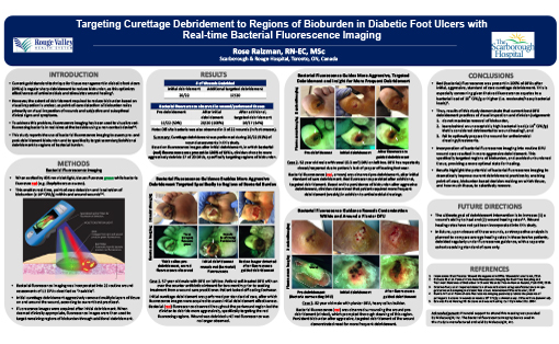
The current gold standard for tissue management in diabetic foot ulcers is regular sharp debridement to reduce bioburden. However, a recent study by Rose Raizman, RN-EC MSc, a wound care clinician, which used bacterial fluorescence imaging with the MolecuLight i:X after initial, aggressive debridement in 20 diabetic foot ulcer wound assessments, found that persistent bacterial red fluorescence was still present in 100% of these debrided diabetic foot ulcers. This led the wound care clinician to perform additional debridement under fluorescence guidance, specifically targeting the regions of remaining bioburden.
Results of this pilot study demonstrated that best current diabetic foot ulcer debridement practices of visual inspection and wound care clinician judgement:
- Do not maximize removal of bioburden.
- Leave behind an unacceptably high bacterial load (≥ 104 CFU/g)2 that is considered detrimental to wound healing.3
- Fail to optimally prepare the wound for antimicrobial dressings/treatments.
As a result, using the MolecuLight i:X has the potential to improve current debridement practices by enabling point of care visualization of bacterial load, to specifically guide removal of bioburdened tissue and avoid un-burdened tissue.