ABSTRACT
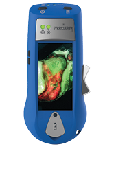
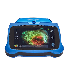
A 64-year-old White woman was admitted to the hospital with complaint of progressive right hip ulceration at the wound site following a total right hip arthroplasty. Initial history and physical examination gave a leading differential diagnosis of pyoderma gangrenosum. Until recently, the exclusion of infection for pyoderma gangrenosum has been largely clinical and supported by cultures/biopsies demonstrating the absence of infection. The MolecuLight i:X (MolecuLight, Toronto, Ontario, Canada) is a novel bedside fluorescent imaging device capable of determining the bacterial burden within a wound in real time. Fluorescent imaging excluded infection at the initial visit, and debridement was avoided. Subsequently, pathergy was avoided as well. The patient was started on topical clobetasol with hypochlorous acid-soaked dressings. She also received 80 mg daily of prednisone and high-dose vitamin D3 (10,000 IU). Recovery was complicated by a deep tunnel along the incisional line at 3 months postdiagnosis, which required slowing of the prednisone taper and the addition of colchicine. Repeat cultures grew Parvimonas, Pseudomonas, and Streptococcus species. Appropriate antibiotics were given. The patient was transitioned from prednisone to adalimumab and started on negative pressure wound therapy. Negative-pressure wound therapy was discontinued at 5 months, and the wound resolved at 6 months.


